Category Archives: Cancer

A possible clue into cancer recurrence


Breast cancer death rates overall have steadily declined since 1989, leading to an increased number of survivors. But while breast cancer survivors are grateful their bodies show no trace of the disease, they still face anxiety. Breast cancer can and does return, sometimes with a vengeance, even after being in remission for several years. The Conversation

By studying the “cannabilistic” tendency of cancer cells, my research team has made some progress in finding out why.

The chances of recurrence and disease outcome vary with cancer subtype. About one-third of patients diagnosed with triple negative breast cancer, the most aggressive subtype, may experience a recurrence in another part of the body. This is called distant recurrence.

It has been difficult, if not impossible, to predict if and when the same cancer will recur – and to stop it. Recurrent disease may arise from just a single cancer cell that survived the initial treatment and became dormant. The dormancy allowed it to hide somewhere in the body, not growing or causing harm for an unpredictable amount of time.

Determining what puts these dormant cells to “sleep” and what provokes them to “wake up” and begin multiplying uncontrollably could lead to important new treatments to prevent a demoralizing secondary cancer diagnosis.

Recently, my research team and I uncovered several clues that might explain what triggers these breast cancer cells to go dormant and then “reawaken.” We showed that cell cannibalism is linked to dormancy.

How do bone stem cells affect breast cancer?

Breast cancer can recur in the breast or in other organs, such as the lungs and bone. Where breast cancer decides to grow depends largely on the microenvironment. This refers to the cells that surround it, including immune cells, cells comprising blood vessels, fibroblasts and the select proteins they produce, among other factors.

Over a century ago, a surgeon named Stephen Paget famously compared the organ-specific prevalence of cancer metastasis to seeds and soil. Because breast cancer often relapses in bones, in this metaphor, which still holds forth today, the bone marrow provides a favorable microenvironment (the “soil”) for dormant breast cancer cells (the “seeds”) to thrive.

Just as seeds need soil to provide an environment for growth, cancer cells need an environment to grow.
From www.shutterstock.com

Thus, a substantial amount of recent work has involved trying to determine the role in cancer dormancy of a special type of cell, called mesenchymal stem cells (MSCs). These are found in bone marrow.

MSCs in bone marrow are highly versatile. They are able to form bone, cartilage and fibrous tissue, as well as cells that support the immune system and formation of blood. They are also known to travel to sites of tissue injury and inflammation, where they aid in healing.

Breast cancer cells readily interact with MSCs if they meet in the bone marrow. They also readily interact if the breast cancer cells recruit them to the site of the primary tumor.

My research team and I recently focused on potential outcomes of these cellular interactions. We found an odd thing happens, which may provide insight into how these breast cancer cells hide for a long time.

In the laboratory setting, we produced breast tumor models containing MSCs. We also re-created the hostile conditions that naturally challenge developing tumors in patients, such as localized nutrient deficits caused by rapid growth of cancer cells and overcrowding.

We discovered that cancer cells under this duress become dormant after eating, or “cannibalizing,” the stem cells.

Our analysis provided compelling data
demonstrating that the cannibalistic breast cancer cells did not form tumors as rapidly as other cancer cells, and sometimes not at all. At the same time, they became highly resistant to chemotherapy and stresses imposed by nutrient deprivation.

Dormant cells are widely linked to recurrence. We hypothesize that cannibalism thus is linked to recurrence.

What is cellular cannibalism, and why is it important in cancer?

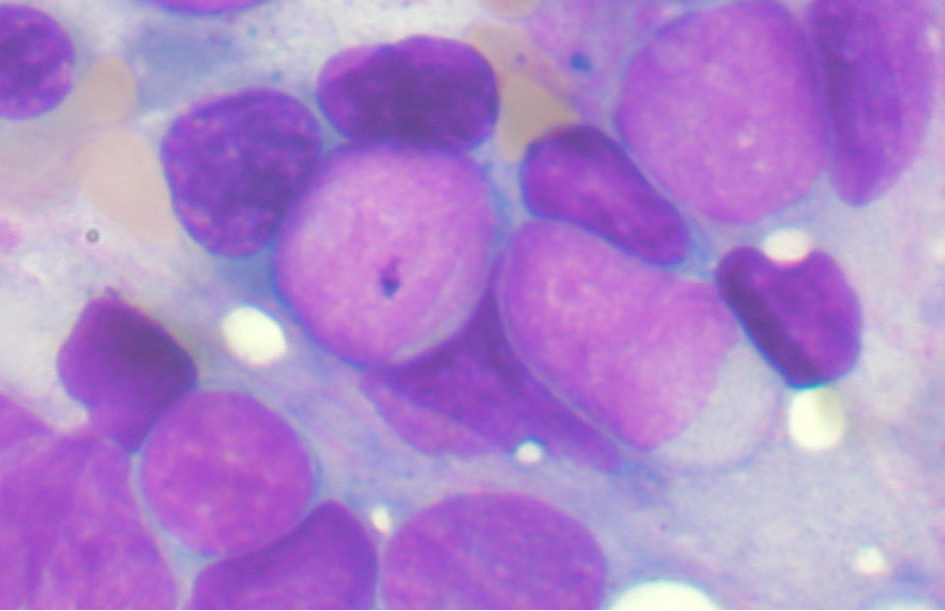
Cellular cannibalism, in general, describes a distinct phenomenon in which one cell engulfs and eliminates neighboring, intact cells.

The percentage of cancer cells that show cannibalistic activity is relatively low, but it does appear to increase in more aggressive tumors.

There are several reasons breast cancer cells would want to eat other cells, including other cancer cells. It provides them with a way to feed when nutrients are in short supply. It also provides them a way to eliminate the very immune cells that naturally stop cancer growth. Cell cannibalism might also allow cancer cells to inherit new genetic information and, therefore, new and advantageous traits.

Notably, in our study, cannibalistic breast cancer cells that ate the stem cells and entered dormancy began to produce an array of specific proteins. Many of these proteins are also secreted by normal cells that have permanently stopped dividing, or senescent cells, and have been collectively termed the senescence-associated secretory phenotype (or SASP). Although cellular senescence is a part of aging, we are now realizing that it is also important for a variety of normal bodily processes, development of embryos and injury repair in adults.

This suggests that although dormant cancer cells do not multiply rapidly or form detectable tumors, they are not necessarily sleeping. Instead, at times they might be actively communicating with each other and their microenvironment through the numerous proteins they manufacture.

Overall, this might be a clever way for dormant cancer cells to “fly under the radar” and, at the same time, modify their microenvironment, making it more suitable for them to grow in the future.

Can cell cannibalism be exploited for diagnosis and treatment?

Although our results are promising, it’s important to be cautious. While there appears to be a strong correlation between cell cannibalism and dormancy, for now we do not know if it is directly linked to cancer recurrence in patients. Studies are underway, however, to corroborate our findings.

Still, the fact that breast cancer cells cannibalize MSCs is intriguing. It provides an important foundation for developing new diagnostic tools and therapies. Indeed, we currently have several ways of applying our recent discoveries.

One exciting idea is to exploit the cannibalistic activity of cancer cells to feed them suicide genes or other toxic agents, using MSCs as a delivery vehicle, like a tumor-seeking missile.

Importantly, MSCs can be easily obtained from the body, expanded to large numbers in the laboratory, and put back into the patient. Indeed, they have already been used safely in clinical trials to treat a variety of diseases due to their ability to aid in tissue repair and regeneration.

A different avenue for drug development would involve keeping dormant cells in a harmless and nondividing state forever. It might also be possible to prevent cancer cells from eating the stem cells in the first place.

In our study, we were able to block cell cannibalism using a drug that targets a specific protein inside cancer cells. With this treatment approach, the cancer might essentially starve to death or be more easily killed by conventional therapies.

Thomas Bartosh, Assistant Professor, College of Medicine, Texas A&M University

This article was originally published on The Conversation. Read the original article.

Ultrasound-activated bubbles could help make cancer drugs more effective and less nasty


Despite extraordinary advances in new drugs and biotechnology, cancer is still one of the leading causes of death worldwide.

In many cases, the problem lies not with the drugs but rather the difficulty in successfully delivering them to the site of a tumour. In healthy tissue there is a regular structure of blood vessels supplying oxygen and nutrients to cells, which divide and grow at a steady rate. In cancerous tumours, however, cells divide and grow in an unregulated way, producing a chaotic vessel structure and regions of tissue with little or no blood supply.

This means when drugs are ingested or injected into the blood stream, they don’t reach all parts of the tumour and there is a high risk of cancer recurring after treatment. On top of this, the pressure inside many tumours prevents a drug from being absorbed from the blood, meaning only a very small fraction of it is actually delivered. The rest of the drug circulates around the body and is eventually absorbed by healthy tissue, often leading to intolerable side effects.

One of the major goals of the research being carried out in the Oxford Institute of Biomedical Engineering (IBME) is to develop new methods for delivering anti-cancer drugs that overcome these barriers. While engineers are perhaps more commonly thought of in the context of large construction projects, we are using precisely the same combination of applied science and problem solving.

Building nanoparticles

There is a formidable series of challenges to address to solve this problem. First, we need to encapsulate the drug to prevent it from interacting with healthy tissue and/or deactivating before reaching the tumour. Second, we need a way to deliver the drug to the tumour to maximise the concentration it receives.

Third, we need a mechanism for releasing the drug on demand once it has built up within the tumour. Fourth, we need to ensure the released drug is evenly spread throughout the tumour. And finally, we need to be able to monitor the treatment from outside the body.

Taking the fight to cancer
Shutterstock

Our team at the IBME has developed a range of new techniques for creating tiny particles into which we can insert drugs with a high degree of precision. And we have tried a variety of methods to make the particles release the drug. These include using materials that are sensitive to the pH change within a tumour and materials that break down upon heating or undergo a phase change (from a solid to a liquid or liquid to a gas).

But one of the most versatile means of triggering drug release is by firing a beam of ultrasonic vibrations at the particles. Widely used as an imaging method, ultrasound can be used from outside the body and, unlike light or heat, can be tightly focused to produce highly localised effects.

In order to produce particles that respond to ultrasound, we have to include in them a gas or a liquid that easily vaporises. When exposed to the ultrasound, the gas/liquid will undergo a rapid expansion and force the drug out of the particle.

Ultrasound activated bubbles

This process generates a pulsating gas or vapour bubble that has several other significant benefits for drug delivery. The motion of the bubble produced by the ultrasound field helps to drive the drug out of the blood vessels and deep into the surrounding tumour. We have shown that bubbles can push drugs up to four times deeper into tissue than they would normally diffuse, sufficient to achieve a uniform spread throughout a tumour.

There is also a growing body of research that shows microbubbles and ultrasound make cancer cells more permeable to drugs, speeding up the rate at which they work and ultimately cell death. The microbubbles’ motion produces a secondary ultrasound signal that can be detected outside the body. This means the location and activity of the particles can be continuously monitored, providing real-time feedback on the progress of the treatment.

Our aim over the next five years is to translate these developments into clinical use. The work will focus on improving the delivery of four classes of drug that have shown enormous potential but that currently struggle to get inside a tumour and/or have unacceptable side effects. By combining our expertise in encapsulation with the use of ultrasound and shockwaves, we hope to create more effective drugs that can be delivered straight to the location of a tumour and monitored with advanced imaging techniques.

This article is adapted from the 2015 IET A. F. Harvey Prize Lecture

The Conversation

Eleanor Stride is Professor of Engineering Science at University of Oxford.

This article was originally published on The Conversation.
Read the original article.

Why do some breast cancers come back?


Cancer is a collection of many hundreds of diseases. The common factor is that once-normal cells have undergone a series of mutations in their genes that has led to uncontrolled growth and an impaired ability to die when they normally should.

Cancers may also spread into other organs, forming secondary cancers, called metastases. When patients die of cancer, it’s usually due to these metastases.

Breast cancer is one of the most common cancers in the Western world, with 15,000 women (and about 70 men) diagnosed each year in Australia. Fortunately, with modern treatments, more than 90% of women with breast cancer go on to have a normal life expectancy, though the side effects of both the cancer and its treatment affect many aspects of their lives.

When detected, cancer can be classified into stages, based on how advanced the disease is in the body.

In breast cancer, the important factors include the aggressiveness of the cells (the grade) and specific proteins that they make. These proteins drive the growth of the tumour cells, including some that bind to female hormones such as oestrogen and growth-promoting proteins such as HER2. Whether a tumour has involved lymph nodes under the arm is also of great importance in assessing its likely potential to spread further.

These markers guide us closely in what drug treatments to consider but also suggest a “prognosis” – that is, how likely the cancer is to be cured or to come back.

So, a patient with breast cancer may undergo surgery to remove the lump and any involved lymph nodes, radiotherapy to try to ensure the cancer does not come back in the breast or lymph nodes nearby, and drug treatments that depend on these markers of aggressiveness. This is done as an “insurance” to increase the chances that the tumour never returns. Scans such as computed tomography (CT scans) are not usually helpful to monitor for recurrence, as small numbers of tumour cells can still be present, but cannot be seen.

Yet for 10% of patients the disease will return – often many years later – and this person is likely to die eventually of cancer. Even though other treatments may shrink the cancer, they cannot get rid of it all together, so unfortunately cure is not possible.

It is assumed that before this recurrence occurs, tiny microscopic nests of cancer cells are lying dormant somewhere in the body. A major quest for cancer researchers has therefore been to find where these cells are hiding and what causes them to wake up and cause secondary cancer.

One intriguing observation has been that in up to 10% of patients previously treated for cancer who are apparently “cancer free”, very careful examination of both blood and bone marrow reveals a few residual cancer cells. This is strongly linked with a more likely chance of cancer coming back.

However, this is not universal. And we know that many supposed cancer cells floating in the bloodstream will in fact be mopped up by the bodies’ immune system or will die “of natural causes”. So, can we better define which are which?

One promising feature under intense scientific scrutiny is the so-called mesenchymal state of the cells. This indicates the cancer cells have changed from looking even less like their cell of origin – in this case, a breast cell – to more primitive cells that can move uninhibited in blood and spread through tissues. This is the same process the body uses in developing embryos and in other situations such as wound healing.

These mesenchymal features allow cancer cells to survive in the toxic environment of the bloodstream, to evade many of our current treatments such as chemotherapy and to set up home in distant organs – the process of metastasis, or secondary cancers.

We still don’t know what causes cancer cells to undergo mesenchymal change (termed epithelial-mesenchymal plasticity or EMP), but understanding it means we are a step closer to developing drugs that can modify or stop the process. It also takes us closer to identifying a biomarker, so we can determine which patient may benefit from these as-yet-undeveloped drugs.

The Conversation

This article was originally published on The Conversation.
Read the original article.

Why is CRISPR the Science Buzzword of Early 2015?


CRISPR isn’t just the cutting edge of genetic modification – it is re-framing our understanding of evolution.

 What is CRISPR?
CRISPR is a DNA sequence that can do something most other genes can’t. It changes based on the experience of the cell it’s written in.  It works because of a natural ability for cells to rewrite their own genetic code, first discovered in 1987. The name CRISPR was coined in 2002, and it stands for “clustered regularly interspaced short palindromic repeats”. They function as a method of inserting recognizable DNA of questionable or dangerous viruses into DNA strands so that the offspring of the cell can recognize what its ancestors have encountered and defeated in the past. By inserting a CRISPR-associated protein into a cell along with a piece of RNA code the cell didn’t write, DNA can be edited.A 2012 breakthrough  involved, in part, the work of Dr. Jennifer A. Doudna. Doudna and the rest of the team at UC Berkley were the first to edit human DNA using CRISPR.  Recently, in March 2015, she warned this new genome-editing technique comes with dangers and ethical quandaries, as new tech often does. Dr. Doudna in a NYT article, she called for a planet-wide moratorium on human DNA editing, to allow humanity time to better understand the complicated subset of issues we all now face.
CRISPR-related tech insn’t only about editing human genes, though. It affects cloning and the reactivation of otherwise extinct species. It isn’t immediately clear what purpose this type of species revival would have without acknowledging the scary, rapidly increasing list of animals that are going extinct because of human activity. Understanding and utilizing species revival could allow humans to undo or reverse some of our environmental wrongs. The technique may be able to revive the long lost wooly mammoth by editing existing elephant DNA to match the mammoth‘s, for instance. Mammoths likely died out due to an inability to adapt to natural climate change which caused lower temperatures in their era, and are a non-politically controversial choice but the implications for future environmentalism are promising.
Each year, mosquitoes are responsible for the largest planetary human death toll. Editing DNA with CRISPR bio-techniques could help control or even wipe out malaria someday. The goal of this controversial tech is to make the mosquito’s immune system susceptible to malaria or make decisions about their breeding based on how susceptible they are to carrying the disease. The controversy around this approach to pest and disease control involves the relatively young research behind Horizontal Gene Transfer, where DNA is passed from one organism to an unrelated species. A gene that interferes with the ability of mosquitoes to reproduce could end up unintentionally cause other organisms to have trouble reproducing. This info is based on the work of , , http://www.biorxiv.org/content/early/2014/12/27/013276
Even more controversial are the startups claiming they can create new life forms, and own the publishing rights. Austen Heinz’ firm is called Cambrian Genomics which grows genetically-controlled and edited plants. The most amazing example is the creation of a rose species that literally glows in the dark. Cambrian is collaborating with the rose’s designer, a company called Glowing Plant, whose projects were eventually banned from kickstarter for violating a rule about owning lifeforms. Eventually, Heinz wants to let customers request and create creatures: http://www.sfgate.com/business/article/Controversial-DNA-startup-wants-to-let-customers-5992426.php#photo-7342819
The final example in an ongoing list of 2015 breakthroughs involving CRISPR is this CRISPR-mediated direct mutation of cancer genes in the mouse liver might be able to combat cancer. It’s the second cancer-related breakthrough in 2015 that affects the immune system, the first was on Cosmos about a week back: Accidental Discovery Could Turn Cancer Cells Into Cancer-Attacking Immune Cells.

Other Related Cosmoso.net articles:

Pre-Darwinian Theory of Heredity Wasn’t Too Far Off

Wooly Mammoth Poised to be the First De-Extincted Animal, Son~!


Jonathan Howard
Jonathan is a freelance writer living in Brooklyn, NY

Angelina Jolie’s surgery got you worried?


Following her 2013 announcement in the op-ed pages of The New York Times that she was having a double mastectomy, US actress Angelina Jolie Pitt has published another piece this week discussing her decision to have her ovaries and fallopian tubes removed to mitigate her high genetic risk of cancer.

Jolie Pitt carries a faulty BRCA1 gene, which predisposes women to developing breast and ovarian cancer. Three women in her family – her mother, aunt and grandmother – were diagnosed with breast or ovarian cancer while still under the age of 60. All three died of their illness.

The publicity surrounding her double mastectomy led to what researchers and the media have dubbed the “Jolie effect”. An Australian study published six months after Jolie Pitt’s disclosure found referrals to familial cancer centres in Victoria more than doubled, and 64% involved people with a high risk of breast cancer. A similar UK study showed that in the year following her May 2013 announcement, referrals to 12 family history clinics increased over twofold.

But ovarian cancer, as you will see, is very different to breast cancer in that it’s very rare. So those of us who work in the field actually hope there’s no Jolie effect in this instance because it’s likely to cause a lot of worry to women who don’t need to be concerned and to divert resources away from those who do.

BRCA and cancer risk

The genes known as BRCA1 and BRCA2 usually help prevent cancers. Everyone has two copies of both but, in some people, one of the copies of either has an error or fault so it doesn’t work properly. The result is a high risk of developing breast and ovarian cancer at younger ages than usual.

The lifetime risk of ovarian cancer for a woman with a faulty BRCA1 gene is about 40% to 60%. This risk increases from her late 30s and continues on an upward trajectory with age. Breast cancer risk is also higher for these women and can be up to 80% depending on family history.

The ovarian cancer risk for a BRCA2 fault is not as high as for BRCA1, at between 15% and 25%.

An estimated one in five ovarian cancers occurring at or before the age of 60 is due to a faulty BRCA gene. But only around 1% to 2% of women carry a faulty BRCA gene. Most women without it have only a 1% risk of developing ovarian cancer and a 10% risk of developing breast cancer.

Other gynaecological cancers, such as cervical or uterine cancer, are not known to be associated with the BRCA genes.

Mitigating risk

The surgery Jolie Pitt has just undergone involved the removal of both her ovaries, as well as fallopian tubes. That’s because evidence suggests cancer can start in the tubes and travel to the ovaries.

Removing both ovaries and tubes of women with a BRCA fault reduces ovarian cancer risk by 90%. The remaining risk is due to cancer cells that may have already travelled to other sites.

It’s important to note that some women with a BRCA fault who have had their ovaries and tubes removed go on to develop what’s called primary peritoneal cancer some years later. This can happen even if the tubes looked normal when they were removed. A cancerous cell may have already spread into the peritoneal cavity before surgery, or cancer could have developed there independently. Cells lining the peritoneum can cause a cancer that looks indistinguishable from ovarian cancer.

Removing both ovaries also has the benefit of reducing breast cancer risk by 50%, likely due to the onset of early menopause. A downside of having this surgery is that it prompts the change of life, or menopause, at a younger age. Most women having their ovaries and tubes removed because of a high ovarian cancer risk do so five to ten years before the age of natural menopause, which is around 50-years-old.

Early menopause can result in health issues such as an increased risk of heart disease and osteoporosis, which can be mitigated by hormone replacement therapy. Because of this, a doctor will advise the woman about whether she should use hormone replacement, which may also help delay or reduce the onset of menopausal symptoms, such as hot flushes, premature ageing of tissues, vaginal thinning (causing sexual discomfort) and decreased libido.

One way to reduce the risk of ovarian cancer is by using the oral contraceptive pill, which can halve your risk with five years of use.

Don’t panic

Of course, it would be far better to have a reliable screening test to detect ovarian cancer at an early, curable stage before women develop symptoms. Sadly, neither of the two tools we have now can do this.

The CA-125 blood test is no longer recommended because it detects cancer at a point when it can no longer be cured. And internal pelvic ultrasounds, which look for abnormalities in the ovary, are not sensitive enough to pick up early changes. Both help diagnose established cancers that would usually be picked up within three months anyway because of symptoms.

Jolie said she had planned to have her ovaries and tubes removed ten years before the youngest woman in her family was diagnosed, but this is not a universal rule for women who carry a BRCA fault. Usually, we use the more blanket approach of surgery around 40 years of age, which is when most women have had their children. Earlier surgery would further increase the risk of problems associated with early menopause.

Women who have had ovarian cancer and are concerned about others in their family should ask their doctor whether the BRCA genes might have played a role in their illness. Those who have a close relative, such as a mother or a sister, who was diagnosed with ovarian cancer while younger than 70 should contact Ovarian Cancer Action (UK), Ovarian Cancer Australia, Ovarian Cancer National Alliance (US) or consult their doctor.

Genetic counselling and testing through a familial cancer centre may be recommended for some. For women who have the faulty BRCA genes, there’s ongoing peer and professional support.

Women who don’t have a close relative with ovarian cancer do not need to seek advice based on the surgery Jolie has just undergone.

Jolie Pitt’s op-ed about her double mastectomy had a positive impact as it galvanised many women to have their risk of breast cancer assessed, including some who needed to be tested for the BRCA mutation. This latest announcement should not have the same effect as far fewer women are at high risk of ovarian cancer.

Acknowledgement: This article was co-authored by Maira Kentwell, senior genetic counsellor and manager of the Department of Genetic Medicine and Familial Cancer Centre, The Royal Melbourne Hospital.

The Conversation

This article was originally published on The Conversation.
Read the original article.

Whether you ‘battle’ cancer or experience a ‘journey’ is an individual choice


The way we talk about illness matters. This is perhaps no more evident than in the many passionate critiques of the metaphor of the “fight” against cancer, which many of us will eventually “lose”.

In the 1970s, Susan Sontag famously exposed the negative implications for patients of this “military rhetoric about cancer”. In 2010, Robert S. Miller listed the military metaphor as one of “eight words and phrases to ban” in cancer care because, despite some finding it useful, many patients detest it. Kate Granger, a doctor with advanced cancer, warned that she would come back to curse anyone who described her as having “lost her brave fight”. She wrote:

I do not want to feel a failure about something beyond my control. I refuse to believe my death will be because I didn’t battle hard enough … After all, cancer has arisen from within my own body, from my own cells. To fight it would be ‘waging a war’ on myself.

Battle metaphors should not be imposed on patients by their families, healthcare professionals or even by well-meaning fund-raising campaigns. Not surprisingly, some official strategies have opted to talk about a patient’s “cancer journey” rather than bellicose metaphors for the patients’ experience. The Cancer Institute of New South Wales discourages the media from talking about the patient’s “fight” against cancer, instead suggesting “journey” as an acceptable alternative.

Angelina Jolie Pitt talked about her own mother “fighting” ovarian cancer for almost a decade before dying aged 56, but used a journey-related metaphor for her own life when she wrote in the New York Times after surgery to remove her ovaries: “I feel at ease with whatever will come, not because I am strong but because this is a part of life. It is nothing to be feared.”

A reinforcing expression

However, recent research we published in BMJ Supportive & Palliative Care shows that we should focus less on what metaphors to ban or promote, and more on how different metaphors work for people with cancer. We analysed a 500,000-word collection of online forum contributions by people with cancer. Using a combination of close textual analysis and computer-aided methods, we identified 2,493 uses of metaphors in the data, including 899 violence metaphors (such as “battle” and “fight”) and 730 journey metaphors. We then considered the implications of each use by looking at its context. There was no simple dichotomy between violence and journey metaphors.

Journey and strength words.
Purple Sherbert Photography

Both types of metaphors can be used to express and reinforce a sense of disempowerment in the experience of illness, which is usually associated with negative emotions. Conversely, both can also be used to express and reinforce a sense of empowerment, usually associated with positive emotions. Empowerment here is to do with the degree of agency that the patient has, where the person actually wants to have that agency.

There is no question that violence metaphors can be detrimental for patients. They can contribute to helplessness and anxiety, for example when patients writing on the online forum say that they feel “attacked” or “invaded” by the cancer, or describe it as a “killer” that “strangles and shocks your soul”. If the battle metaphor is used for the terminal phase of the disease, it can make someone feel like a failure or guilty for not winning.

Yet in our data, the word “fighter” was always used positively to praise oneself or others for being active, determined and optimistic in spite of difficult circumstances. One person explicitly said: “cancer and the fighting of it is something to be proud of.” Amanda Bennett makes this very point in a passionate TED talk about the “exhilarating fight” she and her husband decided to fight together against the cancer of which he eventually died.

In the same way, journey metaphors can be empowering when they are used to express a sense of acceptance, purpose and control, which may even lead to finding some positive aspects of being ill, or when they are used to suggest companionship and solidarity with others – of being “all in it together.”

Journey metaphors do not position the disease as an opponent, and therefore appear to cause no harm. However, things are not quite so simple. For several patients in our data journey metaphors were disempowering. They were used to express feelings of helplessness and frustration, particularly in the face of “navigating” a journey that patients hadn’t chosen to embark on. Another person talked about people with cancer as “passengers” on a journey they could not control.

Metaphors are resources for talking and thinking about one thing in terms of another, and they come in many varieties: the patients in our data also used metaphors to do with sports, fairgrounds, animals, music, machines, and many others. When metaphors work well, they can be enlightening, comforting, and empowering. When they work badly, they can be confusing, disheartening, and disempowering.

No metaphor works in the same way for everyone. And this is particularly the case when it comes to illness. We should be enabled and encouraged to use the metaphors that work best for us. We are currently working on a “metaphor menu” for cancer patients: a selection of quotations from people with cancer that exemplify the widest possible variety of metaphors. We are exploring how this menu can be made available to patients with new diagnoses. As with dishes in a restaurant, different people will find different metaphors more or less appealing, but, ideally, each individual person will be able to recognise or discover one or more metaphors that are helpful for them.

The Conversation

This article was originally published on The Conversation.
Read the original article.

Accidental Discovery Could Turn Cancer Cells Into Cancer-Attacking Immune Cells


Unexpected results are sort of the point of lab experiments. Laboratory studies reveal the unforeseen and if they didn’t, there would seldom be a reason to perform lab studies. It can be problematic when scientists don’t get the results they wanted or thought to expect but other times new data can be the result of the unexpected, and lead to discoveries no one thought to check for in the beginning. Some famous discoveries happened on total  accident throughout scientific history. The latest unintentional discovery might make one of the most aggressive types of cancer more treatable than ever before.

Scientists at Stanford recently discovered a way to force leukemia cells to become mature immune cells do something amazing.  The researchers were actually trying to stabilize cancer cells so they could keep them alive longer in order to study them. The method of keeping the cells alive allowed the cells to develop into immune cells that may one day help the immune system attack cancerous tumor cells!

You can read the study in full at Proceedings of the National Academy of Sciences.

Acute lymphocytic leukemia (ALL) is the name for a particularly rapidly-progressing cancer where the immature cells that should differentiate and become white blood cells or lymphocytes instead become cancerous.  ALL has several classifications based on which kind of lymphocyte (B cell or T cell) the mutated cancer cell originated from.

The scientists were simply investigating a common type of lymphoblastic leukemia, an acute cancer called precursor B cell ALL, aka B-ALL. B-ALL starts as a rogue B cell mutating away from usefulness during an early part of its maturation. The immature cells can’t fully differentiate and become the B cells they were otherwise destined to be. The flawed B cells lack the  transcription factors  required for normal development. Transcription factors are basically proteins that attach themselves to sections of DNA and are then supposed to switch designated genes on or off, depending on the type of transcription factor.  Did you follow that? It’s a bit technical for the layman but most of us understand DNA. Transcription factors are basically a DNA reader than helps the cell decide which part of your DNA it should use to become a specific type of cell.

So, when a transcription factor messes up and activates the wrong section of DNA or doesn’t activate the correct section, it can cause mutations where the cell doesn’t develop or develops poorly. B-ALL is one of the most nasty types of cancer and the  prognosis for victims is not good. The Stanford U team wanted to study this villain but had trouble keeping the cancer cells alive outside of the victims body.

Lead researcher Ravi Majeti reported in the lab’s news release: “We were throwing everything at the cells to help them survive.”

One of the techniques they used to attempt to keep the cancer cells from dying involved exposure to a certain transcription factor. The exposed cells began to grow and change shape, and the new morphology was a type of white blood cell called a macrophage, normally responsible for attacking  damaged, mutated cells or foreign material.

The team recognized the cancerous cells behaved the same as macrophages in various ways such as surrounding and engulfing bacteria. Most notably, the pseudomacrophages from the cancer cells of mice added back into the cancerous mouse did not behave as a cancer cell, and the mice who did not have cancer did not develop cancer after being exposed.

The Stanford researchers believe the newly converted cells are no longer cancerous. Furthermore, they might even help the body’s immune system regroup and attack other, still cancerous cells. It could work because macrophages normally collect DNA tags from abnormal cells they encounter and also mark foreign material so that other cells in the immune system know what to attack. Since the false macrophages were originally cancerous cells, they will, in theory,  already possess the correct signals that recognize the same kind of  cancer.

Now that this principle has been identified as a possible method of treating one cancer, it might open the door to helping the immune system combat other cancers.

Related Cosmoso article: Pre-Darwinian Theory of Heredity Wasn’t Too Far Off

Jonathan Howard
Jonathan is a freelance writer living in Brooklyn, NY